Jesse Plotkin (Messing Lab) and Myles Joyce (Harris Lab) winners in CNS "Visualizing Science" competition
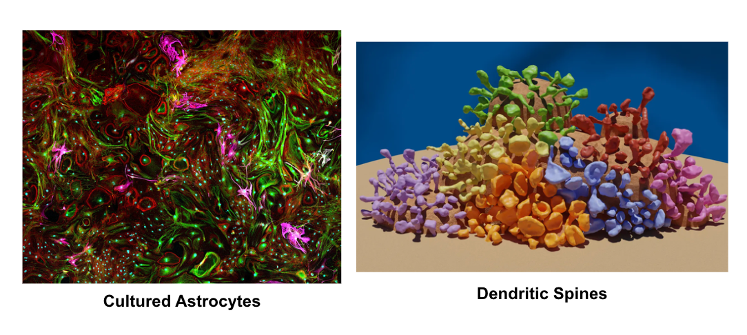
Every year, the College of Natural Sciences invites faculty, staff and students to send in the most striking and fascinating images from their research for the college-wide Visualizing Science competition. The Messing Lab's entrance of cultured astrocytes (image credit Jesse Plotkin) and the Harris' Lab entrance of dendritic spine reconstructions (image credit Myles Joyce) was awarded the Top Prize! Click here to see all the winners!
About the images:
Cultured Astrocytes: Jesse Plotkin and other researchers are conducting a study investigating how star-shaped cells called astrocytes, found in the brain and spinal cord and that are responsible for vital functions in the central nervous system, change over time and whether alcohol exposure has an impact on their development.
The cells pictured here, using confocal microscopy, were from a control sample cultured for two weeks under normal conditions. They were stained using immunohistochemistry with antibodies against s100b (green), a calcium-binding protein expressed predominantly in astrocytes, Glial Fibrillary Acidic Protein (magenta), and Phalloidin (red), a fluorescent tag that binds actin. GFAP and actin are proteins found within the structural cytoskeleton of the cells. The entire image was stitched together by software from many smaller images.
Dendritic Spine Reconstruction: Dendritic spines are specialized protrusions found on the branches of neurons that serve as the main post-synaptic sites for receiving chemical signals in the brain. Highly diverse and dynamic, they change shape throughout life in response to experience and learning. Through countless hours reconstructing rat hippocampal dendrites for his research, neuroscience Ph.D. student Myles Joyce observed how the variety and unique beauty of dendritic spines closely resemble the vibrant biodiversity of coral reefs. In this visual metaphor, each coral polyp is a separate dendritic spine reconstructed from electron micrographs of rat hippocampal neurons. Spines are grouped based on morphology, with different shapes assigned a unique color, highlighting the diversity and complexity of neuronal structure. Just as reefs are built by thousands of distinct yet interdependent organisms, neural connectivity and function arise from the interplay between an orchestra of specialized spines. Nature has a striking tendency to use similar forms and organizational themes across disparate biological systems.